DREAM OCT® VG 200C
VG 200C combines rapid 400 kHz sweeping speed, deep visualization, and ultra-widefield coverage to support advanced OCT/OCTA imaging across retina and anterior segment workflows.
At-a-Glance Technical Snapshot
Core technical specifications of the VG-200C designed for high-speed swept-source OCT imaging, wide retinal coverage, and efficient multimodal workflow.
DREAM Concept: Deep • Rapid • Extensive • Accurate • Multimodal Imaging
The VG-200C integrates high-speed swept-source OCT, deep imaging capability, and widefield performance in one platform for efficient retina and anterior segment workflows.
Deep Imaging
Supports deeper structural visualization for both retina and anterior segment assessment.
Rapid Performance
400 kHz scanning enables fast acquisition and efficient clinical workflow.
Extensive Coverage
Wide scan range and OCTA montage help document larger retinal areas with confidence.
Multimodal Accuracy
Combines SS-OCT, fundus imaging, and automatic alignment in one streamlined system.
Clinical Imaging Applications
The VG-200C is designed to support retina, glaucoma, widefield documentation, and anterior segment imaging within one integrated swept-source OCT workflow.
Retina Imaging
Designed for detailed posterior segment assessment with wide scan capability and strong structural visualization.
Glaucoma Workflow
Supports optic nerve and posterior pole evaluation with high-resolution imaging for routine monitoring.
Widefield Documentation
Helps capture broader retinal regions for efficient clinical review and image documentation.
Anterior Segment Imaging
Extends imaging capability beyond posterior assessment to support cornea and anterior structure evaluation.
Detailed Technical Parameters
Complete technical specifications of the VG-200C for product evaluation, comparison, and procurement reference.
| Category | Specification |
|---|---|
| OCT Technology | SS-OCT |
| Central Wavelength | 1050 nm |
| A-scan Rate | 400 kHz |
| Axial Resolution (Optical) | 3.8 μm |
| Lateral Resolution (Optical) | 10 μm |
| Axial Resolution (Digital) | 2.0 μm |
| Imaging Depth | 12 mm (16.2 mm for AS) |
| Maximum Retina Scan Range | 16 mm |
| Maximum 3D Scan Range (Single Shot) | 15 mm × 12 mm (29 mm × 24 mm with UWF lens) |
| OCTA Montage | 23.5 mm × 17.5 mm (220° with UWF lens) |
| Fundus Imaging Technology | cSSO |
| Light Source | SLD |
| Fundus Wavelength | 830 nm |
| Field of View | 40° × 40° (71.5° × 71.5° with UWF lens) |
| Range of Refractive Compensation | −20D to +20D (−33D to +40D with UWF lens) |
| Tracking Rate | 60 Hz |
| Alignment | Automatic / Electrical |
| Anterior Segment | Supported |
| Posterior Segment | Supported |
| Biometry | Not Supported |
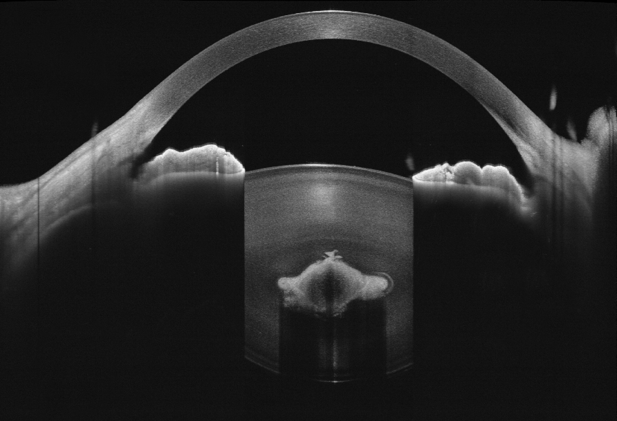
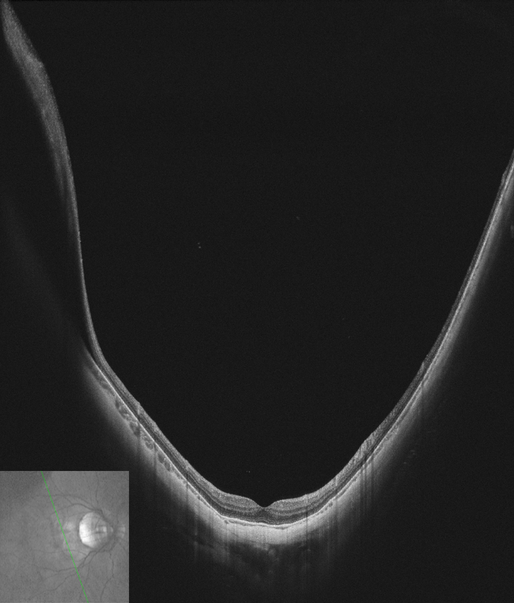
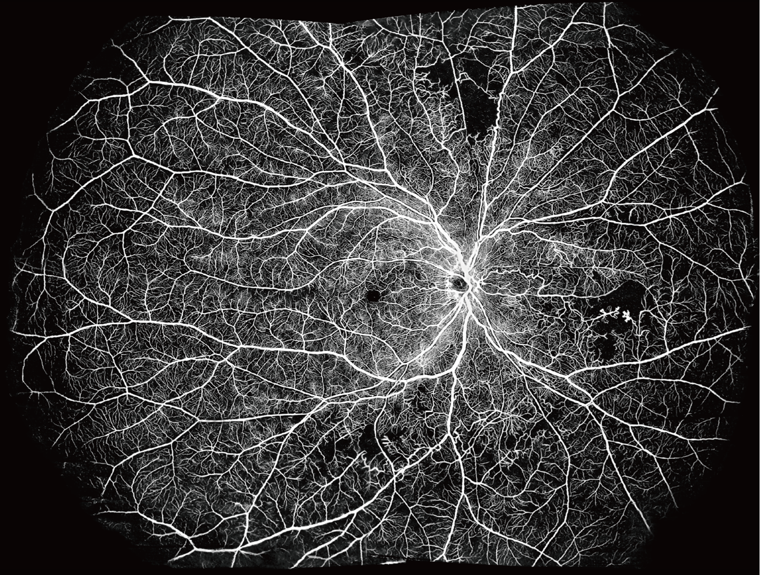
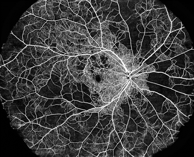
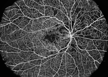
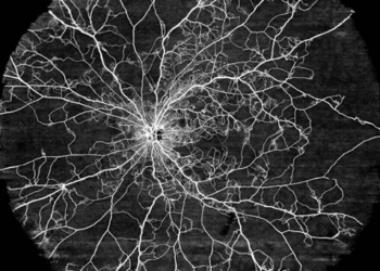
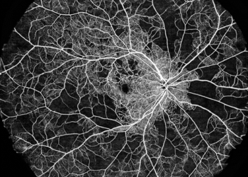
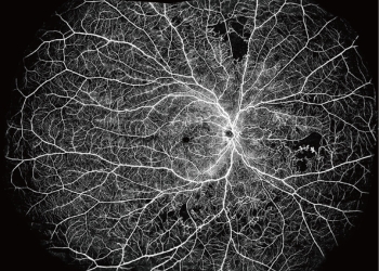
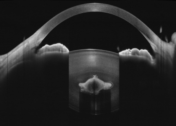
Clinical Imaging Samples
Real clinical imaging examples demonstrating depth, clarity, and ultra-widefield OCTA performance.

Contact Us for Product Inquire
Share your workflow needs and our team will guide you with the right configuration, options, and pricing.