DREAM OCT® VG 200I
VG 200I combines stable 200 kHz swept-source scanning, deep imaging capability, multimodal clinical workflow support, and integrated biometry functionality for retina and anterior segment applications.
At-a-Glance Technical Snapshot
Core technical specifications of the VG 200I designed for reliable swept-source OCT imaging, multimodal fundus integration, biometry support, and efficient clinical workflow across retina and anterior segment applications.
DREAM Concept: Deep • Rapid • Extensive • Accurate • Multimodal Imaging
The VG 200I integrates deep imaging, stable 200 kHz performance, multimodal fundus visualization, and biometry support in one swept-source OCT platform.
Deep Imaging
Supports clear structural visualization for both retina and anterior segment workflows.
- 12 mm OCT imaging depth
- 16.2 mm depth for anterior segment imaging
Reliable 200 kHz Performance
Balanced imaging speed designed for efficient workflow and dependable scan quality in routine clinical use.
- 200 kHz A-scan rate
- Optimized for daily clinical imaging
Wide Retinal Coverage
Supports broader documentation with standard and optional UWF lens-based scan expansion.
- 16 mm retina scan range
- 23.5 × 17.5 mm OCTA montage
Integrated Biometry Workflow
Combines SS-OCT, cSSO fundus imaging, and biometry functionality in one integrated clinical platform.
- Biometry supported
- Automatic / Electrical alignment
Clinical Imaging Applications
The VG 200I is designed to support retina, glaucoma, anterior segment, and biometry workflows within one integrated swept-source OCT imaging platform.
Retina Imaging
Supports high-resolution retinal structure evaluation with broad scan capability for routine and advanced assessment.
- 16 mm maximum retina scan range
- 15 mm × 12 mm maximum 3D scan range
Widefield Documentation
OCTA montage and optional UWF lens support help document larger retinal areas more efficiently.
- 23.5 × 17.5 mm OCTA montage
- Up to 200° with UWF lens
Glaucoma & Optic Nerve Workflow
High optical resolution supports structural analysis for optic nerve and posterior segment review.
- 3.8 μm axial resolution
- 10 μm lateral resolution
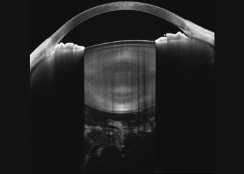
Biometry & Anterior Segment
Integrated anterior segment capability and biometry support extend clinical use beyond standard OCT imaging.
- 16.2 mm AS imaging depth
- Biometry supported
Detailed Technical Parameters
Complete technical specifications structured for detailed evaluation, product comparison, and procurement review.
| Category | Specification |
|---|---|
| OCT Technology | SS-OCT |
| Central Wavelength | 1050 nm |
| A-scan Rate | 200 kHz |
| Axial Resolution (Optical) | 3.8 μm |
| Lateral Resolution (Optical) | 10 μm |
| Axial Resolution (Digital) | 2.0 μm |
| Imaging Depth | 12 mm (16.2 mm for AS) |
| Maximum Retina Scan Range | 16 mm |
| Maximum 3D Scan Range (Single Shot) | 15 mm × 12 mm (26 mm × 21 mm with UWF lens) |
| OCTA Montage | 23.5 mm × 17.5 mm (200° with UWF lens) |
| Fundus Imaging Technology | cSSO |
| Light Source | SLD |
| Fundus Wavelength | 830 nm |
| Field of View | 40° × 40° (60° × 60° with UWF lens) |
| Range of Refractive Compensation | −20D to +20D (−33D to +40D with UWF lens) |
| Tracking Rate | 60 Hz |
| Alignment | Automatic / Electrical |
| Anterior Segment | Supported |
| Posterior Segment | Supported |
| Biometry | Supported |
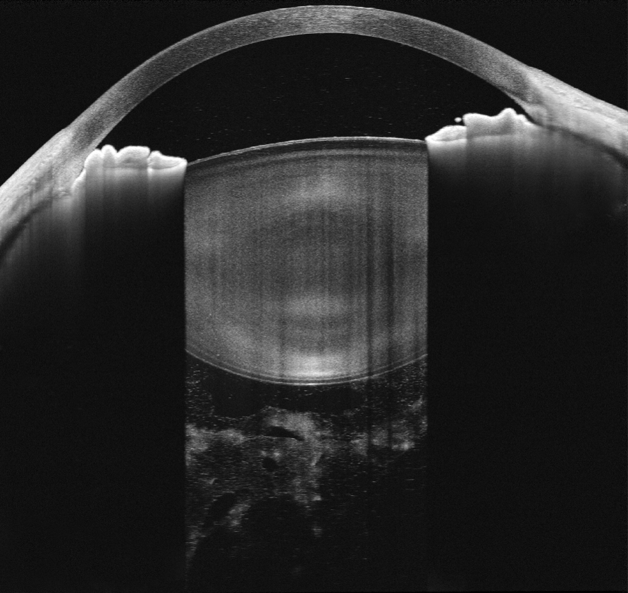
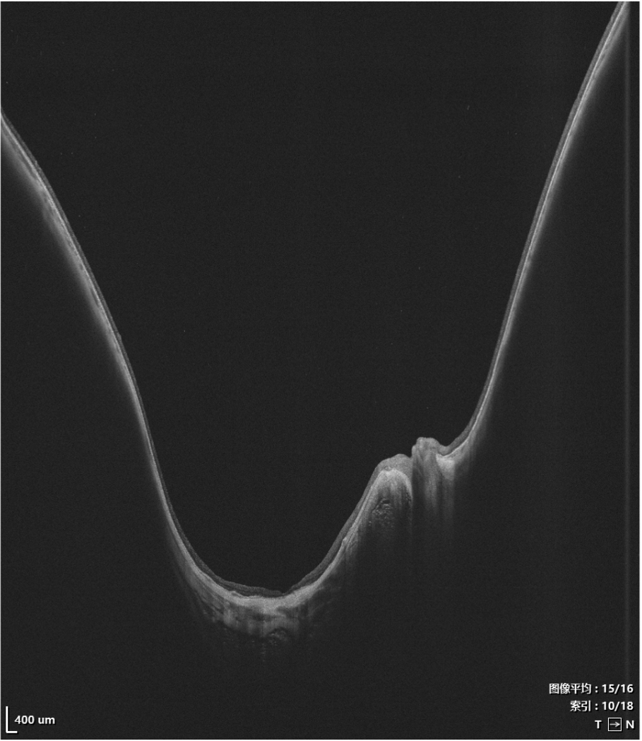
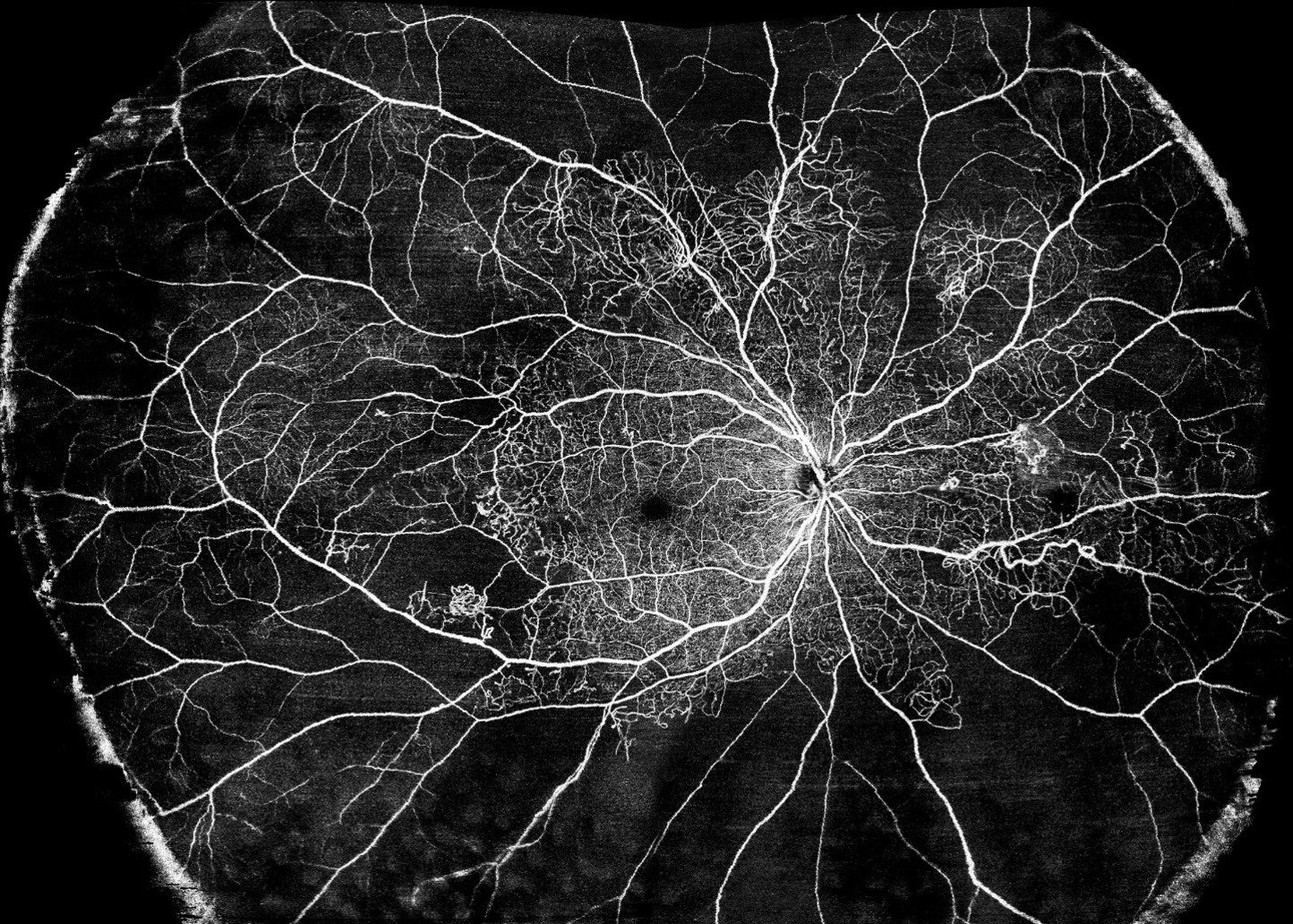
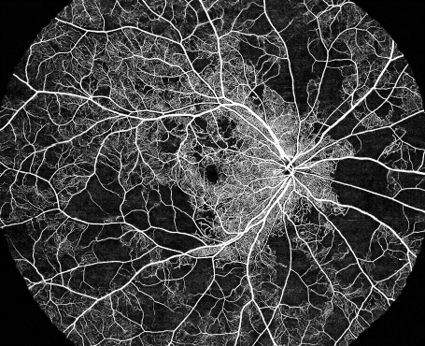
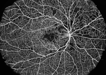
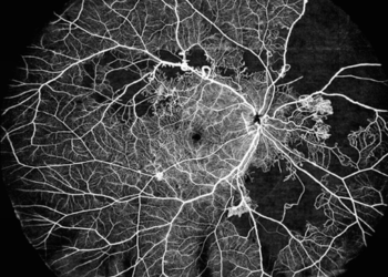
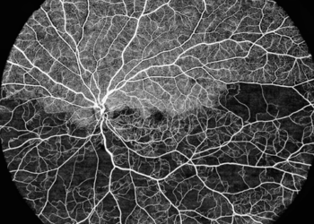
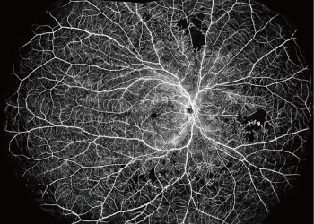
Clinical Imaging Samples
Real clinical imaging examples demonstrating depth, clarity, and ultra-widefield OCTA performance.

Contact Our Team for Product Inquire
Share your requirements (retina / cataract / glaucoma / cornea) and our team will guide you with the right configuration.